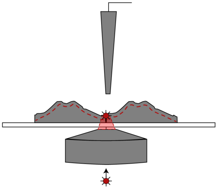
Combining SICM and fluorescence confocal microscopy (SICM-FCM) allows for simultaneous images of topographic detail and fluorescently labelled species at the cell surface. In SICM, the ion current is used to control the vertical position of the nanopipette, meaning that the surface of the cell remains the same distance from the tip of the nanopipette during the scan. In SICM-FCM, the laser is focused just below the tip of the nanopipette meaning that the fluorescent data is spatially aligned with the topographical image.
SICM and fluorescence confocal correlative live imaging of insulin secretion at single granule level in rat insulinoma cells (INS-1E) labelled with NPY-Venus. Released (red arrows), newly released (white arrows) insulin granules and membrane docked dense core vesicles (yellow arrows) are shown. Fluorescently labelled insulin dense core vesiculas are also shown4.
Combining SICM and fluorescence microscopy is a novel technique which allows for direct, label free visualisation of dense core vesicular release at the cell surface.
Combining SICM and fluorescence microscopy is a novel technique which allows for direct, label free visualisation of dense core vesicular release at the cell surface.
Spatially aligned topography, slope, and surface confocal images allows for precise localisation of nanoparticles on the surface cell membrane. This can be seen in the high resolution surface confocal images of 200nm red fluorescent nanoparticles on the surface of lung epithelial cells. A number of protrusions on the topographical image colocalise with red fluorescence spots indicating presence of nanoparticles on the surface. However, there are fluorescent spots (arrowheads) which do not correspond to a topographic protrusion on the surface of the cell, indicating that the nanoparticle has already been internalised. This information could not be deduced from a fluorescence image alone5. At the same time, not all topographic protrusions with dimensions similar to 200 nm sphere colocalise with fluorescence spots, indicating that topography alone also cannot serve as definite proof of nanoparticle presence on the surface.
SICM-FCM is similar to correlative light and electron microscopy (CLEM) as it produces spatially aligned surface and fluorescence images. However, a major advantage of SICM-FCM over CLEM is that it is a live imaging technique which allows dynamic morphological changes at the cell surface to be followed such as the uptake of nanoparticles and viruses3.
Correlative SICM and confocal microscopy
SICM-FCM can be used to visualise internal changes to the cell’s cytoskeleton in real time. As the cells do not need to be fixed, confocal images of the same cell can be taken periodically over time to show cytoskeletal restructuring due to the addition of drugs3.
References
3 - Kolmogorov, V. S. et al. (2021) ‘Mapping mechanical properties of living cells at nanoscale using intrinsic nanopipette–sample force interactions’, Nanoscale, 13(13), pp. 6558–6568. doi: 10.1039/D0NR08349F.
4 - Bednarska, J. et al. (2020) ‘Release of insulin granules by simultaneous, high-speed correlative SICM-FCM’, Journal of Microscopy. doi: 10.1111/jmi.12972.
5 - Shevchuk, A. I. et al. (2008) ‘Imaging single virus particles on the surface of cell membranes by high-resolution scanning surface confocal microscopy’, Biophysical Journal. Elsevier, 94(10), pp. 4089–4094. doi: 10.1529/biophysj.107.112524.


Insulin secretion
Nanoparticle visualisation

ICAPPIC Limited (main office)
+44 (0) 208 383 3080
info@icappic.com
Address: The Fisheries, Mentmore Terrace, London, E8 3PN, United Kingdom

