Patch Clamping on primary cilia
Spatially resolved ion-cannel recordings of primary cilia on mouse IMCD epithelial cells using SICM patch clamping. SICM images show a representation of the spatial positioning of the nanopipette (at the base of the cilium and the extra-cilium membrane). Patch clamp recordings show the single-channel currents for each of the nanopipette position and I/V curves of average single-channel current amplitudes are also shown5.

References
4 - Bhargava, A. et al. (2013) ‘Super-resolution Scanning Patch Clamp Reveals Clustering of Functional Ion Channels in Adult Ventricular Myocyte’, Circulation Research, 112(8), pp. 1112–1120. doi: 10.1161/CIRCRESAHA.111.300445
5 - Torres-Pérez, J. V. et al. (2020) ‘Nanoscale mapping reveals functional differences in ion channels populating the membrane of primary cilia’, Cellular Physiology and Biochemistry, 54(1), pp. 15–26. doi: 10.33594/000000202.


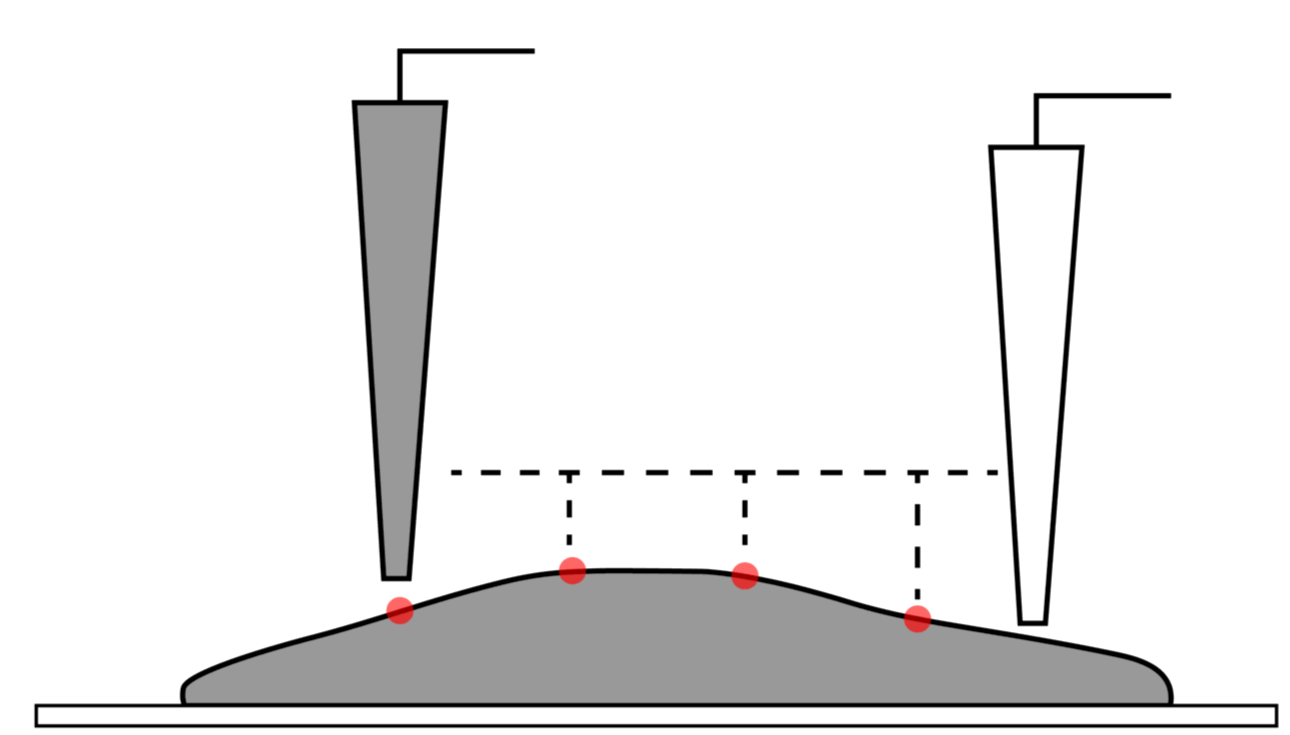
Smart patch clamping combines both SICM and patch clamping to allow accurate positioning of the nanopipette. A topography scan using the SICM capabilities shows the cell’s three dimensional surface nanostructure which is not visible under a light microscope. For ion channel recording, the nanopipette can be positioned accurately. Following contact with the surface, light suction is applied to form a seal and patch-clamping recording can be taken.
The tip of the nanopipette can be broken in a controlled manner to increase the diameter of the tip which may be more suitable for whole-cell patch clamp recordings. This allows for high resolution topography scans to be undertaken using a sharp pipette to identify the region of interest.
Combining SICM and patch clamping allows for high resolution topography scanning of the cell surface allowing the nanopipette to be positioned with nanoscale precision to a specific site of interest such as the T-tubules of cardiomyocytes4.
Patch Clamping on cardiomyocytes


Patch clamping is a technique used to investigate ion channel electrophysiological function in isolated living cells, tissue sections or areas of the cell membrane. However, conventional patch clamp methods do not allow precise selection of the region of interest. Building upon SICM technology, a novel high-resolution scanning patch-clamp system has been developed. This smart patch-clamp protocol enables the study of ion channels in live cells as well as submicrometric cellular structures such as epithelial microvilli.


Smart Patch Clamp

ICAPPIC Limited (main office)
+44 (0) 208 383 3080
info@icappic.com
Address: The Fisheries, Mentmore Terrace, London, E8 3PN, United Kingdom

