
Beta Amyloid Visualisation
SICM topographical images of Human Immunodeficient Virus-like particle formation in Jurkat T-cells2. SICM is able to map virus–surface interactions with nanoscale resolution in physiological conditions.
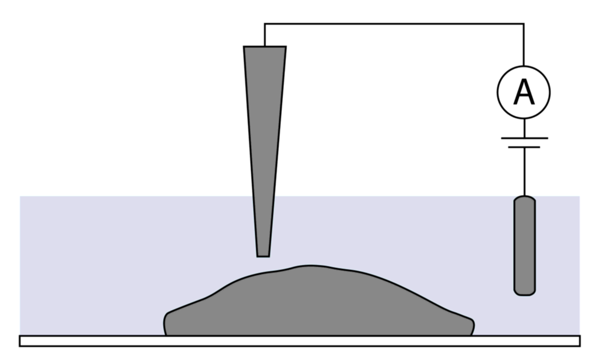
As the nanopipette never touches the sample surface due to the conductance threshold, delicate structures on the surface are not damaged. This allows for continual scanning of the same area to produce sequential images or videos of biological process such as virus assembly.
As the nanopipette never touches the sample surface due to the conductance threshold, delicate structures on the surface are not damaged. This allows for continual scanning of the same area to produce sequential images or videos of biological process such as virus assembly.


A high resolution 3D topography image of cardiomyocytes.

Visualization of vertically protruding mechanosensitive stereocilia of auditory hair cells in the cultured organ of Corti explants. HPICM offers nanoscale resolution of the stereocilia with the smallest measuring ~100 nm or less1 .
ICAPPIC has a patent for HPICM technology. The protocol can be found in Nature Methods1
SICM topographical image of amyloid beta aggregates in DLPC membranes on glass. The nanoscale resolution of SICM allows the accurate imaging of amyloid aggregates offering a unique insight into the mechanics and structures of aggregate formation. Sample courtesy of Dr. Liming Ying, Imperial College London.
References
1 - Novak, P. et al. (2009) ‘Nanoscale live-cell imaging using hopping probe ion conductance microscopy’, Nature Methods, 6(4), pp. 279–281. doi: 10.1038/nmeth.1306.
2 - Bednarska, J. et al. (2020) ‘Rapid formation of human immunodeficiency virus-like particles’, Proceedings of the National Academy of Sciences of the United States of America, 117(35), pp. 21637–21646. doi: 10.1073/pnas.2008156117.
HPICM allows 3D mapping of live cells in physiological conditions without fixing. The technology allows for nanoscale resolution images of delicate structures such as neuronal networks and cilia.

Hopping probe ion conductance microscopy (HPICM) is a form of SICM. HPICM ‘hops’ along the sample surface therefore preventing collisions between the nanopipette and steep structures. This allows topographical imaging of samples with most elaborated surface morphology such as auditory hair cells and neuronal networks. The method can also be tuned adaptively change the digital resolution of the image depending on the topography of the sample. For example, only cell bodies and neuronal processes can be imaged at high resolution, while flat surfaces i.e. empty areas of growth substrate can be imaged at a lower resolution thus accelerating the time in which it takes to obtain the image. Or, vice versa, high resolution can be acquired only on flat regions of a sample avoiding unnecessary oversampling on steep slopes.


Virus assembly and release
3D Mapping

ICAPPIC Limited (main office)
+44 (0) 208 383 3080
info@icappic.com
Address: The Fisheries, Mentmore Terrace, London, E8 3PN, United Kingdom

